Dental Emergencies Welcome | Welcoming New Patients

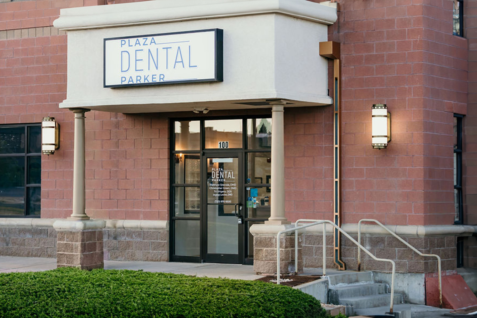
Our Office
10470 S. Progress Way
Suite 100
Parker, CO 80134
Existing Patients: 720-870-9500
New Patients: (720) 640-4406
Visit Us Online

Digital radiography replaces traditional film with electronic sensors and computer processing to capture dental images. Instead of waiting for film to develop, the sensor records X-ray data and converts it into a digital image that appears on a monitor within seconds. This shift from chemical film to pixels has transformed routine dental imaging, making it easier for clinicians to detect issues earlier and explain findings to patients more clearly.
The sensors used in digital systems are compact and designed to fit comfortably in the mouth, producing high-resolution images with greater consistency than older methods. Because the image is produced and stored electronically, it becomes part of the patient’s digital record immediately, simplifying follow-up care and long-term monitoring. For patients, the result is a quicker, smoother experience and a clearer understanding of their oral health.
While the basic goal is the same as traditional X-rays — to reveal structures that are not visible during a clinical exam — digital radiography broadens the possibilities for diagnosis and planning. Dentists can zoom in on suspicious areas, adjust contrast and brightness, and compare images side-by-side to track subtle changes over time. Those enhancements support more informed treatment decisions without adding complexity for the patient.
One of the most important advantages of digital radiography is a significant reduction in radiation exposure compared with conventional film X-rays. Modern digital sensors are more sensitive to X-ray photons, so they require less radiation to produce a diagnostically useful image. This improvement aligns with the long-standing principle of keeping exposure "as low as reasonably achievable" while still obtaining the clinical information needed to deliver excellent care.
Practices using digital imaging also benefit from more precise image capture and fewer retakes, which further limits cumulative exposure over time. In addition, clinics continue to use protective measures such as lead aprons and thyroid shields when appropriate, and they tailor imaging protocols to the needs of children and patients with special considerations. These combined measures help ensure that safety remains a priority during every visit.
Because digital radiography reduces unnecessary repeat imaging, it also supports safer, more predictable diagnostic workflows for patients who require routine monitoring or ongoing treatment. The cumulative effect is a safer imaging environment without compromising diagnostic accuracy — an important factor for patients concerned about radiation and long-term oral health.
Digital images deliver greater clarity and flexibility compared with film. Once an image appears on the screen, the clinician can magnify, crop, or adjust grayscale and contrast to reveal details that might be missed on a flat film. These tools make it easier to detect early decay, assess bone levels around teeth and implants, and spot hairline fractures or root abnormalities that require attention.
Because images are immediately available, clinicians can review findings with patients during the same appointment, using the display to point out areas of concern and outline potential treatment steps. This visual collaboration helps patients understand their condition and the clinical reasoning behind recommendations, which supports informed consent and shared decision-making.
In many cases, digital imaging also reduces the need for additional, follow-up X-rays by capturing exactly what the clinician needs on the first attempt. That efficiency translates into fewer interruptions during appointments and a clearer diagnostic picture from the outset, improving the overall quality of care.
Digital radiography systems connect easily with modern dental practice software, so images are stored directly in the electronic health record alongside notes, treatment plans, and other diagnostic data. This integration supports continuity of care by making historical images accessible whenever they’re needed for comparison, treatment planning, or specialist consultation.
When collaborative care is necessary — for example, orthodontic planning, implant placement, or referral to an oral surgeon — digital images can be shared securely with authorized providers to speed up consultations and reduce delays in treatment. Because the files are electronic, they travel faster than film and retain the same quality when reviewed offsite, which helps specialists provide timely input based on accurate visuals.
Beyond facilitating clinical collaboration, the digital workflow supports reliable recordkeeping and backup protocols, reducing the risk of misplaced films. Secure storage and standardized file formats ensure that images remain usable over many years, helping dentists and patients track long-term trends in oral health with confidence.
There is also an environmental benefit: eliminating chemical processing and film disposal reduces the practice’s ecological footprint. For practices and patients who value sustainability, that’s an additional practical advantage of moving to a digital system.
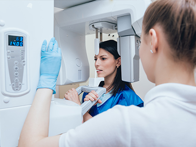
Having digital X-rays taken is quick and straightforward. During the imaging portion of your visit, a small sensor will be positioned inside your mouth for a few seconds while the image is captured. Because exposure times are short and sensors are sensitive, most patients experience minimal discomfort and a brief interruption to the clinical exam. If you have a strong gag reflex or anxiety about in-mouth devices, let the team know — dentists are experienced at working with accommodations to improve comfort.
After each exposure, the images appear on a monitor in the operatory. Your clinician will review the images and explain any findings in real time, showing you the areas of interest and answering questions. This immediate review allows for an efficient visit and ensures you leave with a clear understanding of your diagnostic status and next steps, if any are needed.
Safety precautions are routine: the dental team will use shielding when appropriate and follow established imaging protocols to minimize exposure. For children and expectant patients, imaging recommendations are individualized to balance safety and diagnostic necessity. If additional imaging or a referral is required, your dentist will discuss the reasons and outline what to expect moving forward.
At Plaza Dental Parker, our approach is to use technology thoughtfully — to enhance diagnosis, preserve safety, and keep your experience as straightforward as possible. We combine modern imaging tools with careful clinical judgment so that every image taken has a clear purpose and contributes to better oral health outcomes.
Summary: Digital radiography offers faster imaging, improved diagnostic capability, and reduced radiation exposure compared with film-based methods. It streamlines care through secure digital storage and easier collaboration, while also improving patient communication during appointments. If you’d like to learn more about how digital imaging is used in our office or what to expect during your next visit, please contact us for more information.
Digital radiography uses electronic sensors instead of film to capture X-ray images of the teeth and supporting structures. The sensor records X-ray photons and converts them into a digital image that appears on a monitor within seconds, allowing immediate review. This technology removes chemical processing and integrates images directly into the patient record for convenient access.
Images can be adjusted for brightness, contrast and magnification to reveal subtle findings that might be missed on traditional film. The digital files are stored electronically, which simplifies long-term monitoring and comparison during follow-up visits. Because the workflow is faster and more precise, clinicians can make informed decisions and explain results to patients in real time.
Digital radiography replaces photographic film with compact electronic sensors that are more sensitive to X-rays and produce instant images. Unlike film, digital images do not require chemical developing and can be enhanced, measured and compared on screen to improve diagnostic accuracy. The quicker capture and review process reduces chair time and streamlines clinical workflows.
Digital systems also reduce the need for retakes because images can be evaluated immediately and recaptured if positioning is off. Files remain consistent in quality when shared with specialists or added to an electronic health record. Eliminating film and chemical processing offers environmental benefits as well.
Digital sensors are more efficient than film, so they require less radiation to produce a diagnostically useful image, which helps minimize patient exposure. Practices that use digital imaging typically follow the ALARA principle — keeping exposure as low as reasonably achievable — and use shielding and age-appropriate protocols to protect patients. Fewer retakes from immediate image review further limit cumulative exposure over time.
Imaging recommendations are individualized based on clinical need so patients receive only the images necessary for diagnosis and treatment planning. Routine safety measures such as lead aprons, thyroid protection and collimation are used when appropriate. These combined strategies preserve diagnostic quality while prioritizing patient safety.
Digital radiographs reveal structures that are not visible during a clinical exam, such as interproximal decay, bone loss around teeth and implants, and root abnormalities. The ability to magnify and adjust image contrast helps clinicians identify early-stage lesions, hairline fractures and periapical pathology more reliably. This enhanced visibility supports earlier intervention and more accurate treatment planning.
Digital imaging is also useful for monitoring the progression of periodontal disease, assessing restorative margins and evaluating the fit and placement of dental implants. When diagnostic uncertainty exists, clinicians can compare current and prior images side-by-side to track subtle changes over time. These capabilities contribute to a more thorough and evidence-based approach to patient care.
During a digital X-ray, a small sensor will be positioned in your mouth for a few seconds while the image is captured, and most patients experience minimal discomfort due to short exposure times. The clinician or assistant will position you and the sensor to obtain the necessary views, and the images appear on the operatory monitor almost immediately for review. If you have a strong gag reflex or anxiety about in‑mouth devices, let the team know so they can offer accommodations.
After each exposure, your dentist will review the images with you and explain any findings in real time so you understand the clinical situation and recommended next steps. Standard safety precautions such as shielding are used when appropriate, and imaging protocols are tailored for children and special considerations. This efficient process helps you leave the appointment informed and confident about your diagnostic status.
Digital images provide high-resolution, adjustable views that help clinicians assess anatomy, bone levels and root morphology before performing complex procedures. For implant planning, radiographs assist in evaluating bone quantity and quality and in determining optimal implant position and angulation. In endodontics, magnified digital images reveal root canal anatomy, periapical lesions and subtle fractures that affect treatment strategy.
Because images are immediately available and easily sharable, team members and specialists can collaborate quickly when multidisciplinary input is needed. The digital record also permits precise documentation of preoperative conditions and postoperative monitoring. These advantages contribute to more predictable outcomes and clearer communication with patients about proposed care.
Digital radiographs are stored directly in the electronic health record, where they are organized with your clinical notes, treatment plans and other diagnostic data for secure, long-term access. Storage systems typically include backup protocols and standardized file formats to maintain image quality over time and prevent loss from misplaced film. Access to these records is limited to authorized members of the dental team in accordance with privacy regulations.
When images need to be shared with specialists or outside providers, they are transferred using secure methods to protect patient confidentiality and to ensure that recipients receive high-quality files. Clinics follow established protocols for permissions and audits so that sharing is documented and controlled. This combination of secure storage and regulated sharing improves continuity of care while protecting personal health information.
Digital radiography is used cautiously in children and pregnant patients by tailoring exposure settings and using child‑specific protocols to minimize dose while obtaining necessary diagnostic information. For pediatric patients, sensors and positioning techniques are adapted to ensure comfort and reduce the risk of retakes. Clinicians follow professional guidelines to determine the timing and frequency of imaging based on individual risk and clinical need.
For pregnant patients, routine dental X-rays are generally postponed when possible, but imaging may be performed if it is essential for urgent diagnosis or treatment, with added shielding and careful justification. Dental teams prioritize non‑urgent care and use the lowest effective exposure when imaging is required. Open communication with your provider about pregnancy status helps the team plan safe, appropriate care.
Because digital images appear instantly on a monitor, clinicians can review findings with patients during the same appointment and use visual aids to explain conditions and treatment options. Magnification and contrast adjustments make it easier to point out specific areas of concern, which enhances patient understanding and supports informed decision‑making. Seeing the images alongside an explanation helps demystify the diagnosis and fosters collaborative care.
Immediate image review also shortens the feedback loop, allowing clinicians to answer questions, address concerns and modify treatment plans on the spot. This transparency builds trust and helps patients feel more confident about recommended procedures. Clear visual communication reinforces verbal explanations and written treatment plans for better follow-through.
Choosing a practice that uses digital radiography means benefiting from faster imaging, enhanced diagnostic capability and lower radiation exposure compared with traditional film methods. Digital workflows support precise diagnosis, streamlined recordkeeping and easier collaboration with specialists, which can improve the overall quality and coordination of care. These technical advantages translate into more efficient appointments and clearer explanations tailored to each patient's needs.
At Plaza Dental Parker, our team incorporates modern imaging thoughtfully to support accurate diagnosis and patient education without sacrificing safety. The office pairs digital tools with clinical judgment so every image taken serves a clear diagnostic purpose and contributes to better oral health outcomes.