Dental Emergencies Welcome | Welcoming New Patients

Our Office
10470 S. Progress Way
Suite 100
Parker, CO 80134
Existing Patients: 720-870-9500
New Patients: (720) 640-4406
Visit Us Online

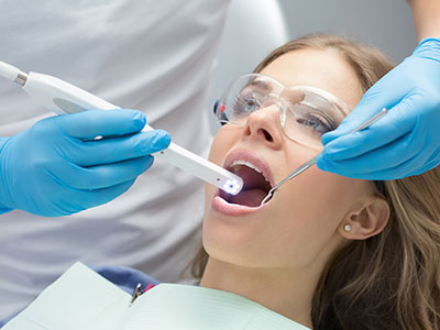
An intraoral camera is a compact, pen-sized imaging device that captures high-resolution, full-color images inside the mouth. Unlike a handheld mirror, it brings tiny details into sharp focus, enabling close-up views of tooth surfaces, gum tissue, restorations, and areas that are otherwise difficult to see. The result is a clear visual record that highlights early wear, cracks, stains, or inflammation before those problems escalate.
These cameras are designed to sit comfortably in the mouth while producing images in real time on a monitor. Patients can watch as the dentist examines each area, helping everyone involved view the same information simultaneously. The camera’s magnification and lighting reveal textures and margins that often escape the naked eye, supporting a more thorough and accurate oral assessment.
Because the images are digital and immediate, the team can compare them to previous photos to track changes over time. That continuity is particularly helpful for monitoring areas prone to recurrence or deterioration, such as margins around crowns, the edges of fillings, and spots of gum recession. Visual documentation turns subjective impressions into objective data that informs ongoing care.
Modern intraoral cameras are engineered to be minimally invasive and fast. They are small enough to navigate the mouth with minimal discomfort, yet powerful enough to deliver crisp images suitable for clinical decision-making. For patients who value clarity and transparency, this technology provides a tangible way to understand oral health as it exists right now.
High-resolution intraoral imaging improves the dentist’s ability to detect problems early, when treatment is typically simpler and outcomes are better. Small fractures, beginning decay, and subtle soft-tissue changes can appear on camera images before they are obvious during a visual exam. Identifying these issues sooner can reduce the need for invasive procedures later and support a more conservative approach to care.
Intraoral photos complement other diagnostic tools, such as digital radiographs and clinical probing, by showing surface details and soft tissue coloration. This layered approach gives clinicians a fuller picture of oral health, helping them distinguish between conditions that appear similar at first glance. When combined with patient history and physical findings, camera images contribute to a more confident and targeted diagnosis.
For complex or ambiguous cases, clear intraoral images make it easier to decide whether further tests or specialist referrals are needed. The level of detail captured helps prioritize interventions and tailor treatment plans to the exact nature of the problem. That precision supports predictable outcomes and helps patients make informed choices about their care.
Because the images are stored digitally, they also serve as a baseline for future comparisons. Tracking subtle changes over months or years can reveal patterns that one-off exams might miss, such as gradual enamel erosion, shifting margins of restorations, or evolving gum disease. This longitudinal view enhances preventive strategies and long-term oral health management.
One of the most practical benefits of intraoral imaging is improved communication between clinician and patient. When a patient can see a clear photo of a concern, explanations about diagnosis and treatment become more concrete and understandable. Instead of relying solely on verbal descriptions, the dental team can point to specific areas in the image and walk patients through what they’re seeing and why it matters.
This visual collaboration builds trust and reduces uncertainty. Patients often feel more comfortable making decisions when they can visually confirm both the issue and the proposed solution. For those who experience dental anxiety, seeing images on a screen from a comfortable distance can make discussions less intimidating than leaning close for a traditional exam.
Intraoral photos also facilitate communication among members of the dental team. Hygienists, specialists, and dentists can review the same images and align their assessments, ensuring a consistent approach to care. That shared understanding helps avoid miscommunication and streamlines the planning process for restorative or cosmetic procedures.
Finally, images can be used as an educational tool to demonstrate home-care needs. For example, close-up photos of plaque accumulation or early gingival inflammation can motivate patients to improve brushing and flossing techniques, making preventive advice more immediate and actionable.
Images captured with an intraoral camera are saved directly into the patient’s digital record, creating a permanent visual history. These records support continuity of care by allowing clinicians to review past conditions, reference prior treatments, and document healing after procedures. Secure, organized photo libraries make it easy to retrieve relevant images when planning follow-up care.
When coordination with a dental laboratory or a specialist is necessary, these images are invaluable. Clear photos help technicians fabricate crowns, veneers, and other restorations that better match the patient’s existing anatomy and shade. Similarly, specialists receiving high-quality images before a referral can prepare more effectively, reducing the need for repeat imaging or extended chair time.
Digital imaging also streamlines clinical workflows. Because photos are captured instantly and stored electronically, they can be incorporated into notes, used in treatment planning software, or referenced during intra-office consultations. This efficiency enhances the patient experience by minimizing delays and ensuring that decisions are based on accurate, up-to-date visuals.
Privacy and security remain central to how these images are managed. Clinical images are handled as part of the patient’s protected health record, stored and accessed according to applicable privacy standards. Patients can be reassured that their visual records are treated with the same confidentiality as other clinical information.
An intraoral camera exam is quick, noninvasive, and designed with patient comfort in mind. During a routine check or targeted evaluation, the dentist or hygienist will gently position the camera to capture the areas of interest. The procedure typically takes only a few minutes, and patients may be invited to watch the images on a screen as they’re taken or to review them afterward with the clinician.
Hygiene protocols are followed rigorously: disposable covers and sterilization procedures protect patients and staff, ensuring that camera use meets clinical infection-control standards. Because the device does not emit radiation, it can be used frequently as needed to document progress, verify the fit of restorations, or monitor healing after treatment.
Some patients appreciate the immediate feedback the camera provides; others prefer to review images privately with the dentist. Either way, clinicians adapt their communication to each patient’s comfort level. For patients who experience gag reflex sensitivity or jaw tightness, the small camera size and gentle technique help minimize discomfort during image capture.
Overall, intraoral imaging enhances the standard dental exam by adding clarity and documentation without adding risk. It supports accurate diagnosis, better patient understanding, and coordinated care, making routine visits more informative and efficient for everyone involved.
At Plaza Dental Parker, we use intraoral camera technology to support clear communication, precise diagnosis, and careful documentation throughout a patient’s course of care. If you’d like to learn more about how intraoral imaging is used during exams and treatment planning, please contact us for more information.
An intraoral camera is a small, pen-sized imaging device that captures high-resolution, full-color photos and video inside the mouth. The camera uses LED lighting and optical magnification to reveal surface details on teeth and soft tissues that are difficult to see with the naked eye. Images are transmitted in real time to a monitor so the clinician and patient can view the same visuals simultaneously.
The device is designed to be minimally invasive and easy to maneuver, allowing targeted shots of hard-to-reach areas such as interproximal contacts and restoration margins. Because the images are digital, they can be saved directly into the patient record for comparison over time. This immediate visual feedback supports clearer diagnosis and discussion during the visit.
High-resolution intraoral images reveal subtle surface changes such as early enamel breakdown, hairline fractures, and margin discrepancies around restorations that may not be obvious during a routine visual exam. These surface details complement other diagnostic tools, allowing clinicians to correlate what they see on camera with findings from clinical probing and digital radiographs. Together, these data points produce a more complete diagnostic picture and reduce the chance of overlooking developing problems.
Having clear, objective photos also helps prioritize interventions and tailor treatment plans to the exact nature of a problem. Images make it easier to document baseline conditions, track progression, and verify outcomes after treatment. That level of precision supports conservative care when appropriate and helps clinicians make more confident recommendations.
An intraoral camera exam is noninvasive and does not expose patients to radiation, making it safe for repeated use as needed. The device is small and rounded to minimize gag reflex and jaw discomfort, and clinicians use gentle techniques to obtain clear images. Standard infection-control measures such as disposable sleeves and sterilization protocols are followed to protect patients and staff.
Most patients find the process quick and unobtrusive; imaging of the full mouth typically takes only a few minutes during a hygiene visit or targeted evaluation. Clinicians adapt their approach to each patient’s needs, offering the option to view images on-screen or review them privately with the dentist. This flexibility helps reduce anxiety while ensuring accurate documentation.
Intraoral images are saved digitally into the patient's electronic health record, creating a secure visual history that clinicians can access for follow-up visits. These images are managed according to applicable privacy and health record standards and are treated as part of the protected clinical record. Secure storage and controlled access help ensure that imaging data remain confidential and available only to authorized team members.
Digital storage also makes it easy to retrieve prior images for comparison, share visuals with a specialist when a referral is needed, or provide photographic documentation to support laboratory communication. Proper organization of image libraries streamlines clinical workflows and improves continuity of care without compromising patient privacy.
Yes. Intraoral cameras excel at showing surface signs of early decay, such as subtle enamel discoloration or cavitation, and can reveal hairline fractures that often go unnoticed during a cursory visual exam. The magnification and lighting emphasize textural changes and marginal irregularities around restorations that may indicate recurrent decay or structural compromise. Detecting these issues early enables more conservative treatment options and can prevent more extensive procedures later.
It is important to note that intraoral imaging complements other diagnostic methods rather than replacing them; radiographs and clinical tests remain essential for assessing subsurface decay and the full extent of structural damage. When findings are ambiguous, clinicians use the combined information from images, X-rays, and clinical evaluation to form a definitive diagnosis and treatment plan.
Visuals make explanations more concrete by allowing patients to see exactly what the clinician sees, which clarifies the nature of a problem and the reasons behind recommended treatments. When clinicians point to a photo and describe the concern, patients gain a clearer understanding of urgency, expected outcomes, and home-care needs. This shared visual reference builds trust and supports informed decision-making.
In clinical practice, images can also be used to illustrate preventive priorities, such as areas with plaque buildup or early gum inflammation, turning abstract advice into actionable steps. By showing progress with follow-up photos, clinicians can reinforce positive behavior and demonstrate the effectiveness of recommended care. At Plaza Dental Parker, we use intraoral imaging to improve communication and involve patients directly in their oral health plan.
No. Intraoral cameras provide detailed surface imagery but cannot visualize internal tooth structure, root anatomy, or bone levels the way radiographs do. X-rays, clinical probing, and other diagnostic tests remain necessary for assessing subsurface decay, periodontal bone loss, and endodontic conditions. The camera adds complementary surface information that enhances interpretation of those other findings.
Using multiple diagnostic modalities together gives clinicians a layered understanding of oral health and reduces the risk of missed pathology. In practice, intraoral imaging streamlines diagnosis and planning by documenting visible conditions that correlate with radiographic and clinical data.
Photographs from an intraoral camera provide precise visual references for shade selection, margin evaluation, and anatomy matching when planning crowns, veneers, and other restorations. High-quality images sent to a dental laboratory or used during in-office design discussions help technicians and clinicians achieve better esthetic and functional outcomes. Visual documentation also assists in verifying fit and finish at try-in appointments.
For cosmetic cases, intraoral photos allow clinicians to demonstrate pre-treatment conditions and plan smile changes with greater specificity. Images support consistent communication among the dentist, dental team, and laboratory, which reduces revisions and improves predictability for restorative and cosmetic procedures.
A typical intraoral imaging session is brief and straightforward: the clinician will gently position the camera to photograph areas of interest while you remain seated in the dental chair. You may be invited to watch the images on a monitor as they are captured or to review them privately with the dentist afterward. The process usually adds only a few minutes to a routine exam or hygiene visit.
Clinicians follow infection-control protocols and use disposable sleeves or sterilized barriers for each patient to ensure safety. If you have sensitivity, a strong gag reflex, or limited mouth opening, the team will adjust technique and positioning to maximize comfort while obtaining diagnostic-quality images.
Because intraoral photos are saved in the digital record, clinicians can compare images from different visits to detect subtle changes such as gradual enamel wear, shifting restoration margins, or evolving gum recession. This longitudinal view makes it easier to spot trends that one-time exams might not reveal and supports early intervention when small changes are detected. Regular photographic documentation contributes to prevention-focused care and long-term planning.
Clinicians use these comparisons to tailor maintenance intervals, evaluate the success of treatments, and decide when a referral or additional testing is warranted. Plaza Dental Parker incorporates intraoral imaging into follow-up care to track progress and ensure treatment goals are being met.